axon terminal diagram
Learn vocabulary terms and more with flashcards games and other study tools. Start studying Axon Terminal Diagram.
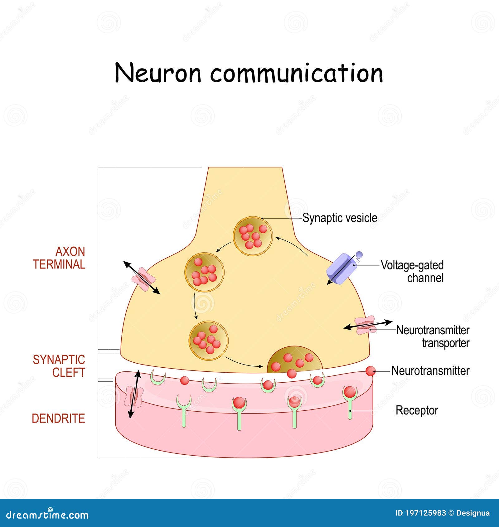
Chemical Synapse Structure Neuron Communication Stock Vector Illustration Of Anatomy Biology 197125983
At this point each axon of the motor neuron will divide into branches called axon terminals.

. The diagram below shows some of the events that occur at a. Figure 15 Diagram of neuron. Define the term axon terminal.
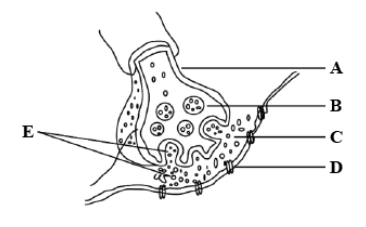
YOU MIGHT ALSO LIKE. E axon terminal E cell body Which of the following structures is labeled C in the diagram. This diagram is most important in our Neet exam and board exam.
The end of the axon has terminals. And are often branched. Axon terminals are button-like or club-like structures at the end of an axon.
The part of the sarcolemma muscle plasma membrane that lies beneath the axon terminalsnerve endings is called motor end plate. The neuron carries messages to other neurons via an axon which is often myelinated to increase the speed of the message. A dendrites B axon terminal C peripheral process D trigger zone E cell body E E This structure.
How to Draw Axon Terminal and Synapse Diagram. A single short process that extends from the cell body and then splits into two branches in opposite directions. One branch travels to the peripheral nervous system PNS.
Synapse Tiny fluid-filled gap from axon terminal of one neuron to the dendrites of another Neurotransmitter A chemical messenger that travels across the synapse from one neuron to. The nervous framework is a profoundly mind boggling part of a creature that facilitates its Q. Axon Axon is a tube-like structure that functions by carrying an electrical impulse from the cell body to the axon terminals for passing the impulse to another neuron.
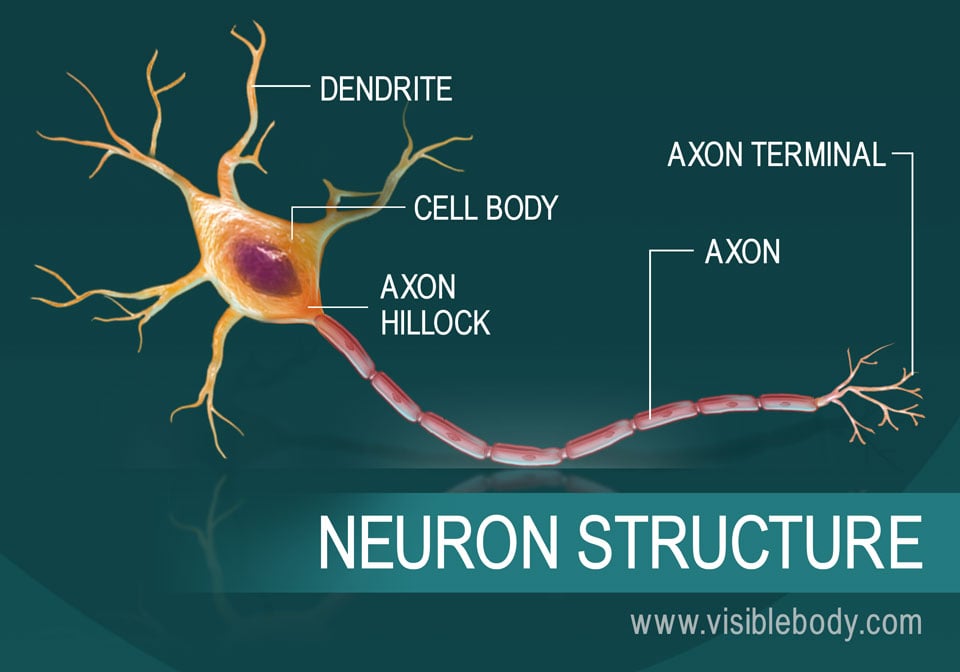
They are often enlarged compared to the rest of the axon. Each axon may also possess side branches. An axon is a cable that transmits messages away from the cell body or soma towards the dendrites of other neurons or the sensory receptors of other types of cells to.
Axon terminal Diagram Quizlet Expert solutions Log in Sign up Axon terminal Flashcards Learn Test Match Created by Darby_Erb Terms in this set 21 1. Towards the end of the axon terminal closest to the muscle fiber the tip of the axon. 2 cell body 3 nucleus 4 nucleolus 5.
Axons may be myelinated by oligodendrocytes in the central nervous system CNS and by Schwann cells in the peripheral nervous system PNS or may be unmyelinated.
Axon Wikipedia

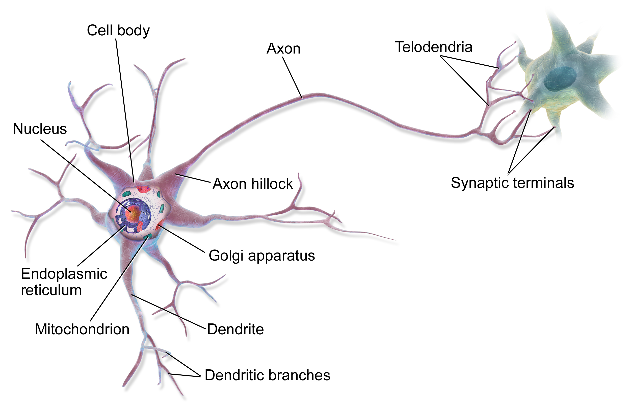
What Is A Neuron Diagrams Types Function And More

Axon Structure And Function Nerve Cell Mcat Content
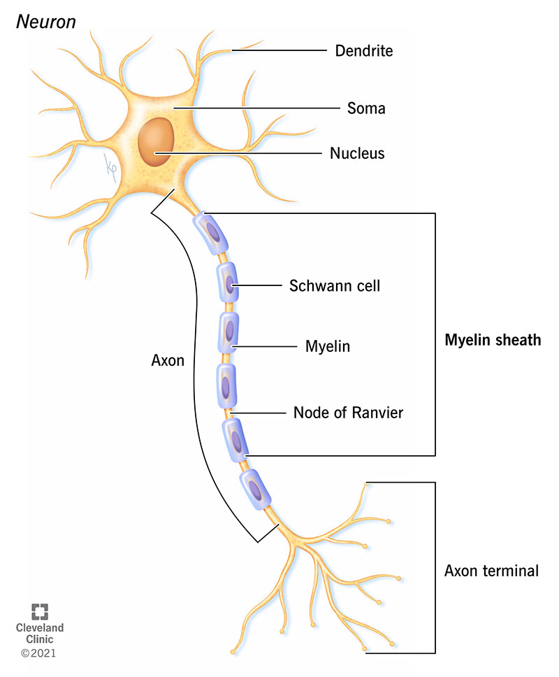
Myelin Sheath What It Is Purpose Function

Schematic Diagram Of Vesicle Fusion And Recycling At The Axon Download High Quality Scientific Diagram
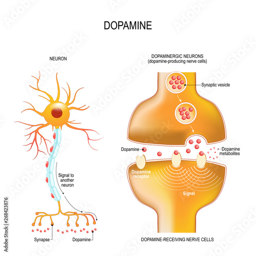
Dopamine Closeup Presynaptic Axon Terminal Synaptic Cleft And Dopamine Receiving Nerve And Dopamine Producing Cells Stock Vektorgrafik Adobe Stock
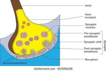
Diagram Showing Axon Terminal Synapse Stock Vector Royalty Free 2076906328 Shutterstock

A Diagram Showing Axon Terminal And Synapse Is Given Identify Correctly At Least Two Of A D

Neuron Diagram
In The Following Diagram Showing Axon Terminal And Class 11 Biology Cbse

Schematic Representation Of A Synapse Showing The Circulation Of Download Scientific Diagram

Diagram Of A Synapse Showing Neurotransmitters Stored In Synaptic Vesicles Inside The Axon Ter Neurotransmisores Anatomia Del Cerebro Humano Biologia Avanzada

Dopamine Vector Illustration Labeled Diagram With Its Action And Pathways Stock Vector Vector And Low Budget Royalty Free Image Pic Esy 057866925 Agefotostock

Axon Terminal Diagra Diagram Quizlet

A B C Ii Iii I

Axon Terminal Definition And Examples Biology Online Dictionary
Neurons